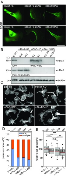
Fig. 8.
Formation of multiple fronts and defects in polarization and migration in mDia1- and/or -3-deficient B16-F1 cells. (A) Subcellular localization of EGFP-mDia1 and -3 variants in B16-F1 cells migrating on laminin. While full-length (FL) mDia1 and -3 were cytosolic and largely excluded from protrusive fronts, constitutively active mDia1-FL-2xAla (M1182A and F1195A), mDia1ΔDAD, mDia3-FL-2xAla (M1057A and F1170A), and mDia3ΔDAD strongly accumulated in the rear cortex. (Scale bar, 20 µm.) (B) Immunoblotting of individual and combined mDia1 and mDia3 KO clones as indicated. GAPDH: loading control. Numbers below respective lanes indicate relative changes of mDia1 and -3 expression levels normalized to GAPDH. (C) Polarization defects and formation of multiple fronts in respective cell types, as revealed by phalloidin staining. Note increasingly pronounced multiple-front phenotypes in single versus double (mDia1/3) KO cells. (Scale bar, 10 µm.) (D) Quantification of protrusive fronts (one versus multiple) from images as shown in C. Error bars, standard error of the mean from at least six independent experiments, n, cell number. (E) Quantification of random migration of respective cell types on laminin. Boxes include 50% and whiskers 80% of all measurements; dots represent the fifth/95th percentile. n.s., not significant; **P < 0.01; ***P < 0.001 by Mann–Whitney rank sum test. n, number of tracked cells.

