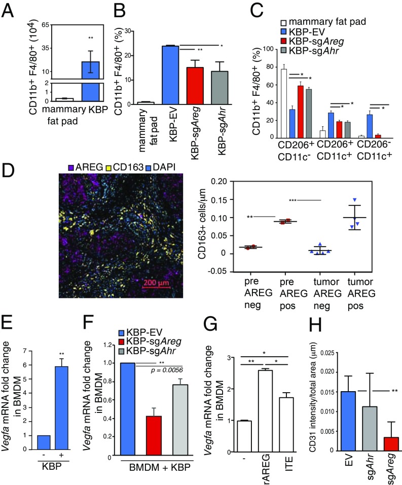
Fig. 4.
AhR−AREG axis influences the number and function of TAM. (A) Absolute numbers of CD11b+ F4/80+ cells in normal mammary fat pad from wild-type virgin and nulliparous mouse females and KBP tumor-bearing female mice (n ≥ 5 per group). (B) Percentages of CD11b+ F4/80+ cells in the mammary fat pad and EV, sgAhr, or sgAreg KBP tumor-bearing mice (n ≥ 5 per group). (C) Percentages of the indicated subpopulations of CD11b+ F4/80+ macrophages in the mammary fat pad and EV, sgAhr, or sgAreg KBP tumor-bearing mice (n ≥ 5 per group). (D) (Left) Representative immunohistochemistry staining of CD163 (surface marker for human macrophages) and AREG staining in a human primary BRCA1-mutated BC. Nuclei were stained with DAPI. (Right) Total count of CD163-positive (CD163+) macrophages in AREG positive (AREG+) or negative (AREG−) areas in reduction mammoplastic (pre) or tumor tissues from BRCA1 mutant carriers. See Materials and Methods and SI Appendix, Fig. S4D for additional details. (E) Vegfa mRNA levels in BMDM cultured alone (−), or after coculture with KBP cells (+) for 24 h (n = 3 per group). (F) Vegfa mRNA levels in BMDM after coculture with KBP cells expressing EV, sgAhr, or sgAreg for 24 h (n = 3 per group). (G) Vegfa mRNA levels in BMDM that were left untreated (−) or treated with rAREG (50 ng/mL) or ITE (10 μM) for 24 h (n = 3 per group). (H) Quantification of CD31 staining intensity as an indicator of angiogenesis in EV, sgAhr, or sgAreg tumors (n ≥ 5 per group). *P ≤ 0.05, **P ≤ 0.01, ***P ≤ 0.001.

