Fig. 2.
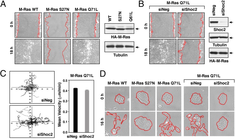
Shoc2 is required for M-Ras–driven collective migration of MCF10A cells. (A) Confluent monolayers of MCF10A cells stably expressing the indicated HA-M-Ras proteins were wounded, and images taken at 0 and 18 h after wounding. HA-M-Ras and tubulin levels are shown. (B) M-RasQ71L–expressing MCF10A cells were transfected with control (siNeg) or Shoc2 siRNAs before wounding. Images were taken at 0 and 18 h after wounding. Lysates were also examined for Shoc2, HA-M-RasQ71L, and tubulin levels. (C) M-RasQ71L–expressing MCF10A cells transfected with control or Shoc2 siRNAs were plated at low density on collagen-coated surfaces, and isolated cells were tracked for their direction and velocity of movement over a 12-h period. (D) Serum-starved clusters of MCF10A cells stably expressing the indicated proteins were induced to scatter by the addition of growth media. Shown are images taken at 0 and 16 h after scatter induction. Red lines indicate free cell edges in A, B, and D.

