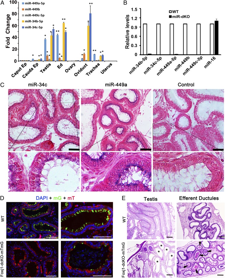
Fig. 2.
miR-34b/c and -449a/b/c are abundantly expressed in the ciliated cells of EDs and mice with ciliated cell-specific knockout of the five miRNAs phenocopy the global double miRNA cluster knockout mice. (A) qPCR analyses on miR-34b/c and -449a/b/c expression levels in the caput and cauda epididymis (Ep), testis, EDs, ovary, oviduct, trachea, and uterus. U6 snRNA was used as an internal control, and the fold changes were calculated against levels in the caput epididymis. Data are presented as mean ± SEM (n = 3). *P < 0.05; **P < 0.01 (Student t test). (B) qPCR analyses on miR-34b/c and -449a/b/c expression levels in WT and miR-dKO EDs. miR-16 was used as an internal control. Data are presented as mean ± SEM (n = 3). (C) In situ hybridization results showing that both miR-34c and -449a are localized to the ciliated cells in the efferent ductal epithelium. Specific hybridization signals are blue. (Scale bars: 50 μm.) (D) Fluorescent microscopic images showing cross-sections of EDs in control (Foxj1-Cre;Rosa26mTmG+/Tg) and Foxj1-dcKO-mTmG (Foxj1-Cre;Rosa26mTmG+/Tg;miR-34b/c−/−;miR-449lox/lox) mice. Expression of membrane-bound EGFP (mG) is indicative of Cre-expressing ciliated cells. The mG-positive ciliated cells are far fewer in Foxj1-dcKO-mTmG EDs than in controls. (Scale bars: 100 μm.) (E) H&E-stained paraffin sections of testes and EDs in WT (Foxj1-Cre;Rosa26mTmG+/Tg) and Foxj1-dcKO-mTmG (Foxj1-Cre;Rosa26mTmG+/Tg;miR-34b/c−/−;miR-449lox/lox) mice. Ciliated cell-specific conditional knockout (Foxj1-dcKO) males appear to phenocopy the global knockout (miR-dKO) males because both display efferent ductal occlusion and sperm agglutination (arrows), as well as disrupted spermatogenesis due to dilated seminiferous tubules (asterisks). (Scale bars: 100 μm.)

