Fig. 3.
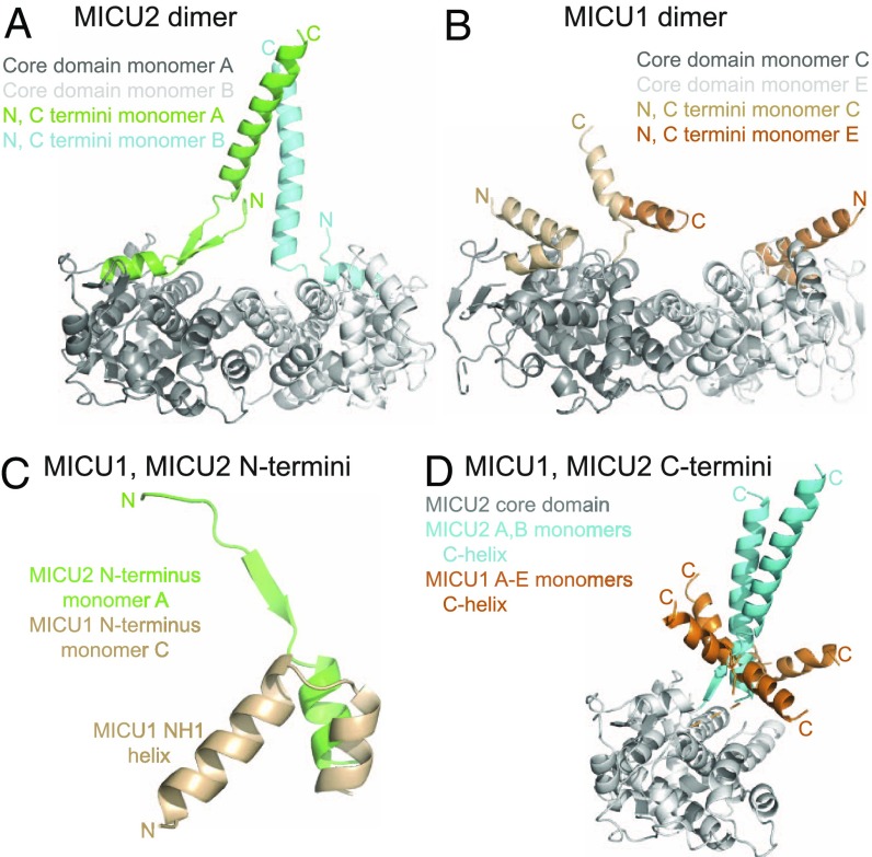
The N termini and C termini differentiate MICU2 from MICU1. (A) The MICU2 dimer (6EAZ) is shown with the N and C termini colored for emphasis, labeled N and C, respectively. (B) The MICU1 dimer (4NSC, C and E monomers) is shown with the N and C termini colored for emphasis. The orientation shown is the same as for the MICU2 dimer in A. (C) The MICU1 and MICU2 N termini are shown highlighting that the MICU1 NH1 helix folds back (toward the core domain), while the MICU2 corresponding region does not. Note that in MICU1 the amino acid sequence immediately preceding NH1 helix is the lysine-rich segment specific for MICU1 which is indispensable for function. (D) MICU1 monomers from the 4NSC hexamer and MICU2 monomers from the 6EAZ dimer are superimposed. The core domain of MICU2 monomer A is shown in gray for reference. The C-helices of the five monomers of MICU1 are colored in orange (note the sixth was not resolved in 4NSC), and the two monomers of MICU2 are colored in cyan.

