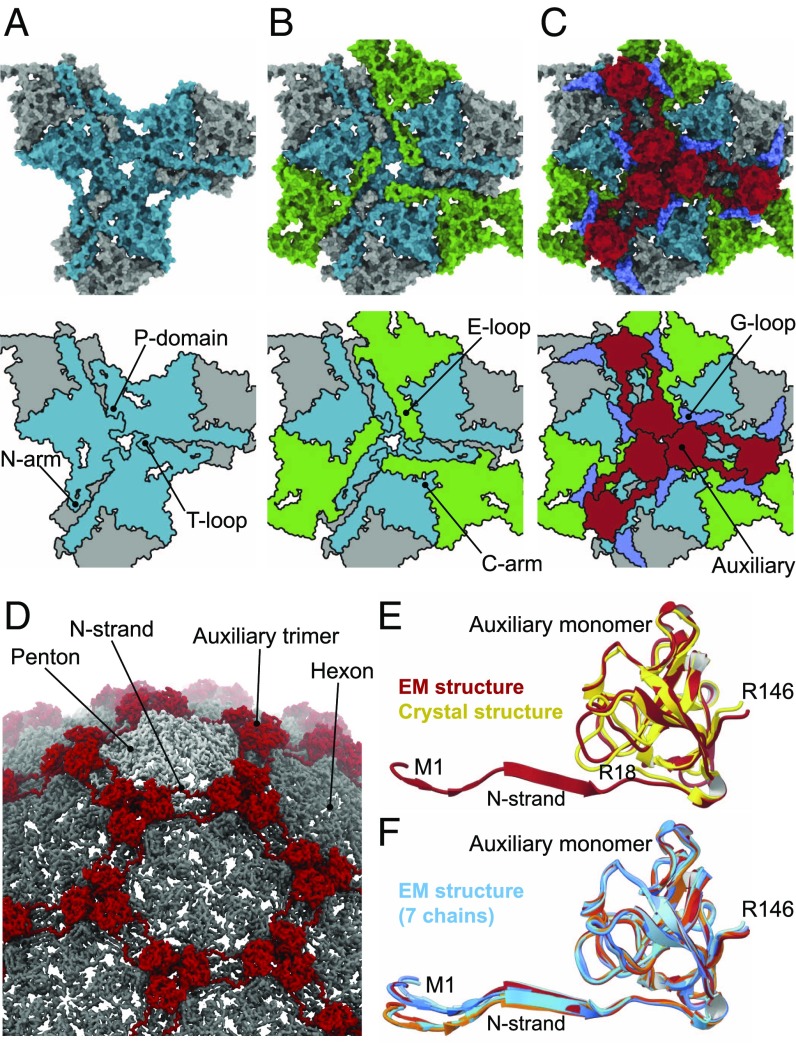
Fig. 3.
The multilayered nature of subunit interactions. (A) Innermost layer of the capsid wall featuring six capsid protein chains centered on a threefold symmetry axis, shown in gray and cyan. (B) A middle layer featuring E loops from three further subunits (green) extending toward the central axis and overlaying the P domains of subunits in the innermost layer. (C) Upper layer containing auxiliary protein trimers (red), and major capsid protein G loops (blue) from the internal layer. Space-filling representations in A–C, Top are shown along with schematic diagrams (Bottom). (D) Segment of the expanded capsid map with annotated features. (E) Overlay of the auxiliary protein monomer from the cryo-EM reconstruction (red, this work) and crystal structure (yellow, PDB ID code 6BL5). (F) Overlay of the seven auxiliary protein subunits constituting the icosahedral asymmetric unit.

