Fig. 4.
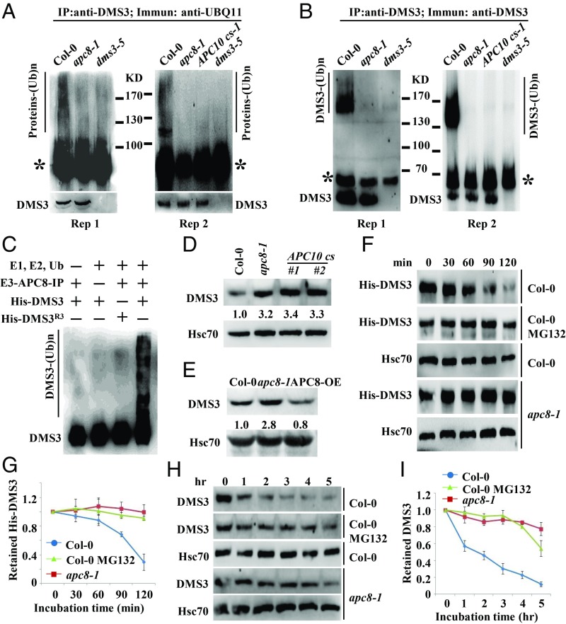
DMS3 is ubiquitinated and degraded by APC/C in vivo and in vitro. (A) Detection of ubiquitination levels of DMS3 immunoprecipitates (IP) in Col-0, dms3-5, APC10 cs-1, and apc8-1. DMS3 immunoprecipitates were immunoblotted with anti-UBQ11 to detect Ub-protein conjugates. The protein standards were indicated. The asterisk indicates the heavy chain of anti-DMS3. (Bottom) Anti-DMS3 blot to indicate the DMS3 levels in each genotype. Two biological replicates are shown. (B) Detection of DMS3 ubiquitination in Col-0, dms3-5, and apc8-1. Anti-DMS3 immunoprecipitates were immunoblotted with anti-DMS3 to detect Ub-DMS3 conjugates. The protein standards were indicated. The asterisk indicates the heavy chain of anti-DMS3. The bottom band indicates the DMS3 level in each genotype. Two biological replicates are shown. (C) APC/C-mediated ubiquitination of DMS3 in vitro. Reactions were carried out with the different combinations of E1, E2, Ub, E3 (anti-GFP immunoprecipitates from APC8-YFP transgenic plants) and the substrates His-DMS3 or His-DMS3R3. The reaction was stopped, and proteins were detected by Western blotting using an anti-DMS3 antibody. The smeared bands indicate ubiquitinated DMS3, while the bottom band indicates nonubiquitinated DMS3. Western blotting was used to determine DMS3 levels in seedlings of Col-0, apc8-1, and cs lines of APC10 (D) or APC8-OE (E). Hsc70 was the loading control. The numbers indicate the average abundance of DMS3 from three biological replicates. (F) Decay rate of His-DMS3 in a cell-free protein decay assay. His-DMS3 was incubated with equal amounts of cell lysate of Col-0 or apc8-1 with or without 50 μM MG132 for the indicated time. The levels of His-DMS3 were determined with anti-His antibody. Hsc70 was used as the loading control. (G) Statistical analysis of the decay rate of His-DMS3 in F. The levels of His-DMS3 were plotted versus the level at 0 min. Data are presented as mean ± SE (n = 3). (H) Decay rate of DMS3 in Col-0 or apc8-1. Total proteins from Col-0 and apc8-1 were extracted and incubated with or without 50 μM MG132 for indicated time. Levels of DMS3 were determined with anti-DMS3. Hsc70 was the loading control. (I) Statistical analysis of decay rate of DMS3 in H. Data are presented as mean ± SE (n = 3).

