
Figure 3. Structural analyses of HA-GCN4IL-sfGFP.
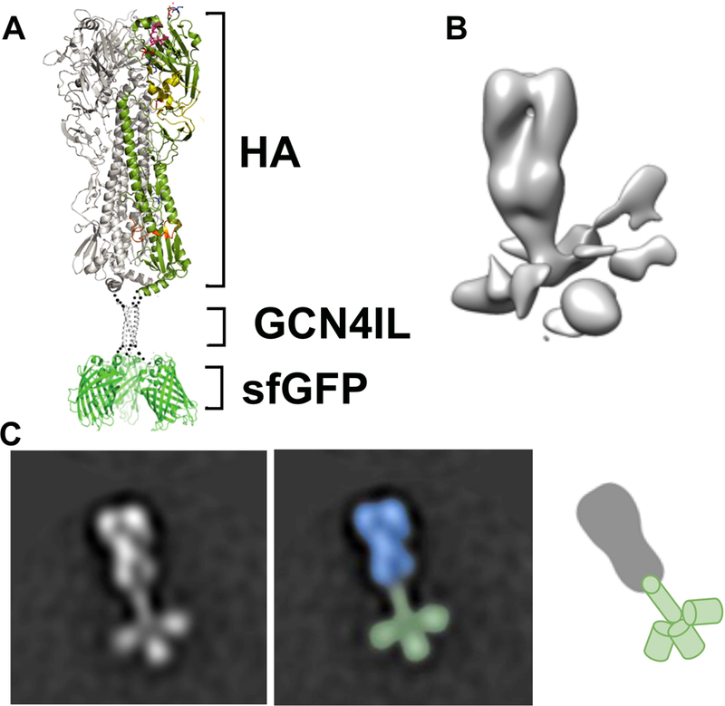
(A) Ribbon representation of the HA-sfGFP trimer. A single protomer in the HA section of the fusion protein is colored by domain. (B) Negative stain 3D reconstruction of the trimeric HA-sfGFP fusion protein. The HA trimer is well ordered while the C-terminally fused sGFP appears as irregular densities at the base of the trimer likely due to flexibility. (C) Negative stain 2D class average showing the 3D structure of the trimeric HA-sfGFP. The distinct HA, GCN4IL and sfGFP structures can be seen resembling the layout as shown in the ribbon structure.