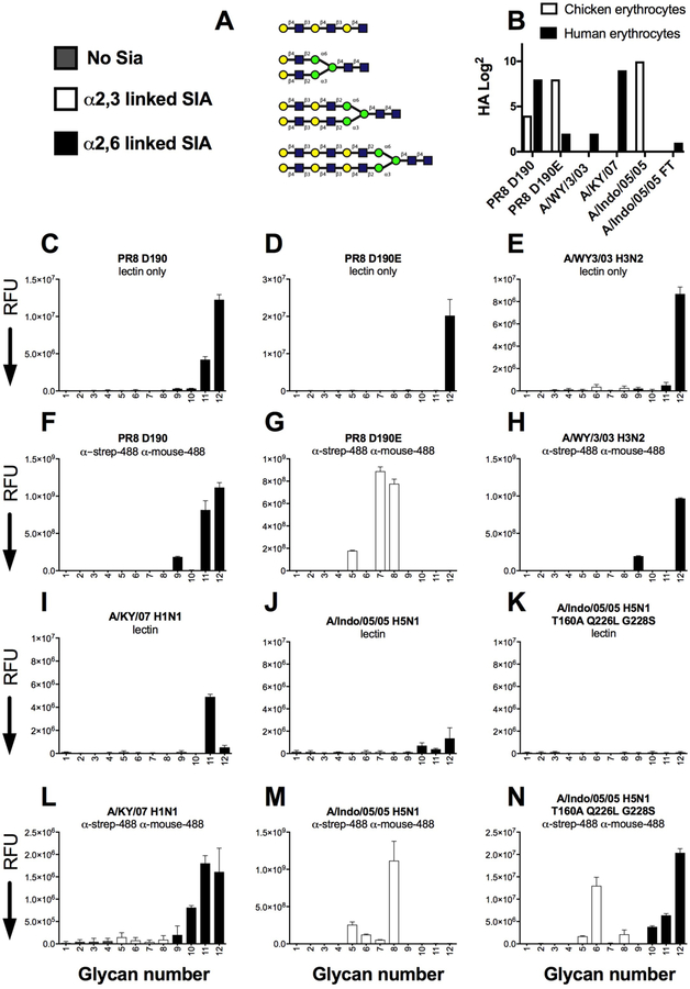
Figure 5. Receptor specificity of different HA as lectins and multivalent complexes on a N-glycan array.

(A) Glycans imprinted on this array, depicted are the unsialylated controls that are all sialylated with either 2,3 linked and 2,6 linked sialic acids. (B) Hemaglutination assay with precomplex HA proteins toward chicken and human erythrocytes. (C–E and I–K) indicated HA proteins as lectins only. (F–H and L–N) indicated HA proteins precomplexed with α-strep and α-mouse abs both labeled with Alexa-488. The mean signal and standard error were calculated from six independent replicates of glycans printed on the array. The data shown is a representative of two independent assays. α2–3 linked sialosides are shown in white bars (glycans 5 to 8 on the x axis) and α2–6 linked sialosides in black (glycans 9 to 12). Glycans 1 to 4 are non-sialylated controls.
