
Figure 7: Presentation of N-glycans capped with α2,3 linked sialic acids on a glass slide lacks complexity for multivalent interaction with an HA trimer.
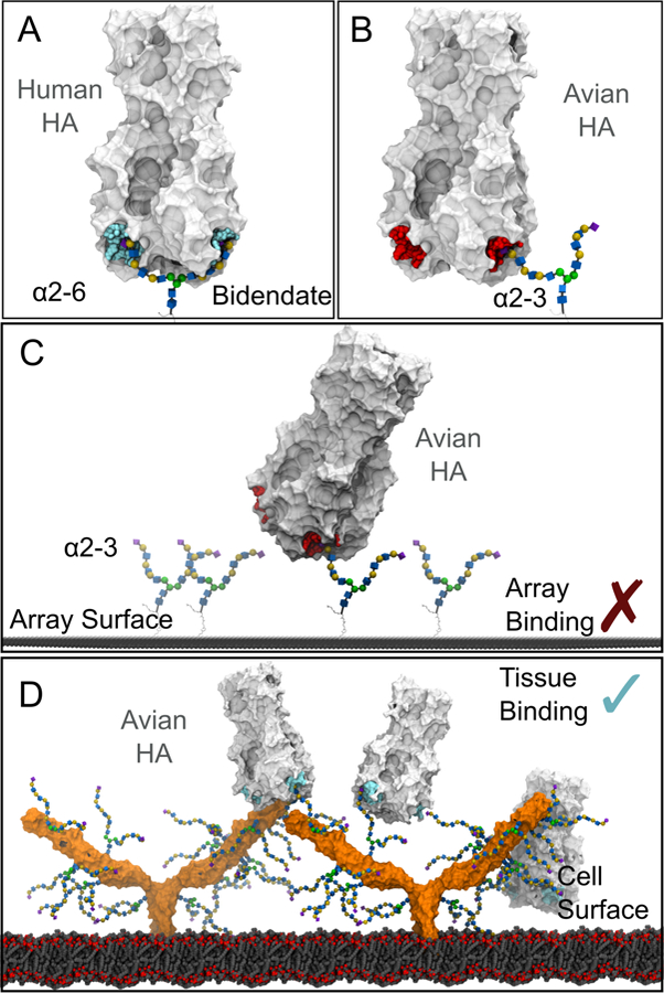
The HA binding sites are colored cyan (to indicate multivalent binding) or red (no multivalent binding). (A) Bidentate binding of a HA to an α2–6 capped, branched N-glycan with three LacNAc repeats on each branch. (B) Bidentate binding is not possible between the equivalent glycan capped with α2–3 linked sialic acid and a HA, due to the different orientation of the sialic acid. (C) On a slide surface glycans capped with α2–3 linked sialic acid must be spaced correctly to form multivalent interactions with a HA, and no recruitment of the glycans is possible. (D) Multivalent binding of avian HA to glycans capped with α2–3 linked sialic acid may be possible on a cell surface, due to the higher density of glycans on cell surface glycoproteins (example shown is ICAM-1, with glycans attached to N-glycosylation sites). HA forms a multivalent interaction with glycans present on one ICAM-1 molecule, or between two different ICAM-1 molecules.