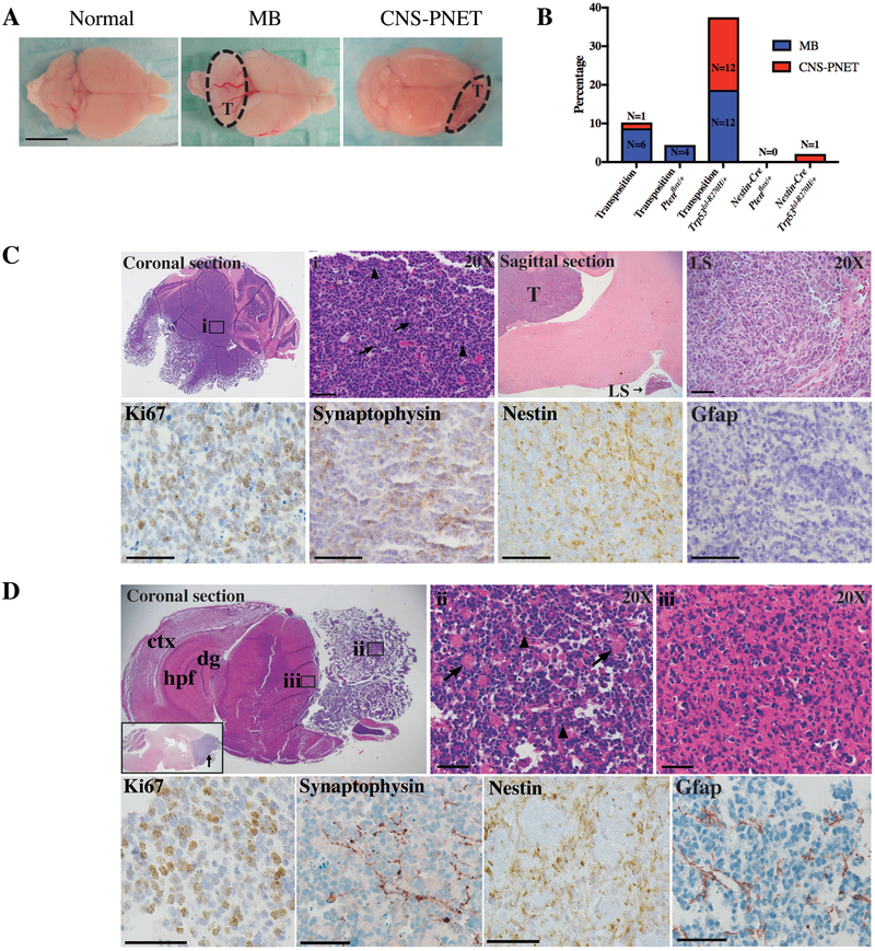
Figure 1.
SB-induced medulloblastoma and CNS-PNET resemble human tumors.
A, Macroscopic images of normal brain and brains with SB-induced cerebellar medulloblastoma and CNS-PNET in the cerebral cortex and olfactory bulbs. T=tumor. B, Medulloblastoma and CNS-PNET frequency across genetic backgrounds. C, Upper panels: medulloblastoma H&E. i, Rosettes (arrows), mitotic nuclei (arrowheads). Primary medulloblastoma (T) with leptomeningeal spread (LS). Lower panels: medulloblastoma IHC. D, Upper panels: CNS-PNET H&E. Cerebral cortex (ctx), hippocampal formation (hpf), dentate gyrus (dg). Inset: CNS-PNET sagittal section, olfactory bulb (arrow). ii, Bulk tumor with rosette formations (arrows) and mitotic nuclei (arrowheads). iii, Tumor cell parenchyma infiltration. Lower panels: CNS-PNET IHC. Scale bars = 50 μm.

