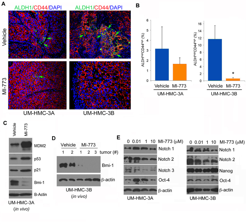
Figure 6.
Effect of MDM2/p53 binding inhibition by MI-773 on cancer stem cells in UM-HMC-3B cells in-vivo. A, Immunofluorescence analysis of UM-HMC-3A and UM-HMC-3B tumors for ALDH and CD44. Images were taken at 200X. B, Graph depicting the percentage of ALDHhighCD44high cells in MI-773 treated UM-HMC-3A and UM-HMC-3B tumors. After 6 days of treatment, the tumors were resected and digested into a single cells suspension. The cells were then stained and FACS analyzed. A t-test was used to test for significance. Error bars indicate the standard deviation. C, Western blot analysis of tissue lysate from UM-HMC-3A vehicle and MI-773 treated tumors. D, Western blot analysis of tissue lysates from xenograft tumor generated with UM-HMC-3B cells that were treated with MI-773 (3 independent tumors) or control (2 independent tumors). E, Western blot analyses of stem cell transcription factors (i.e. Notch-1, Notch-2, Notch-3, Oct-4 and Nanog) in UM-HMC-3A and UM-HMC-3B exposed to 0–10 µM MI-773 for 24 hours.

