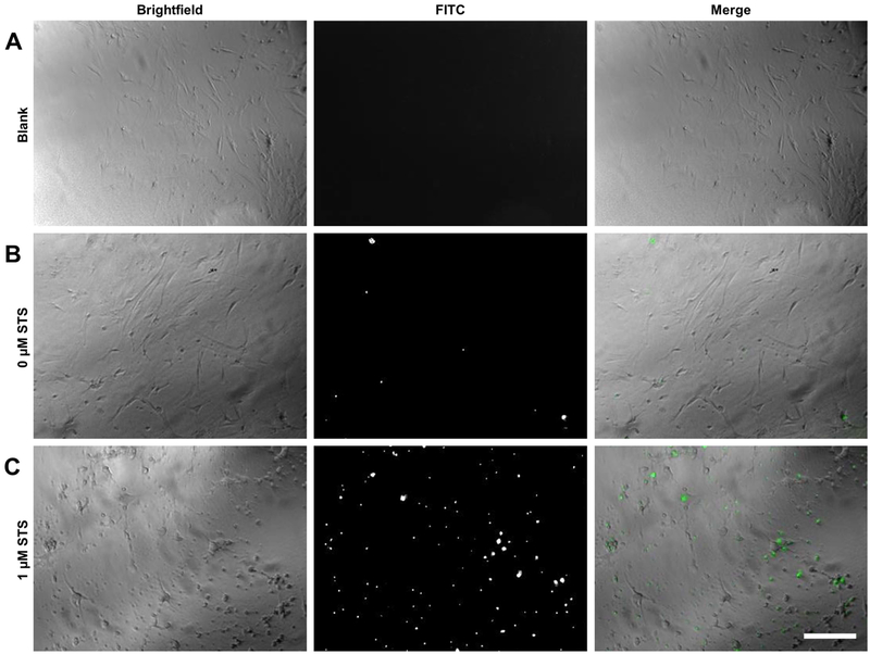
Figure 3.
Fluorescent microscopy of cells treated with IncuCyte® Caspase-3/7 Green Reagent. C57BL/6J MLFs were seeded into microplates at 35,000 cells/mL and treated with either vehicle only (A), IncuCyte® Caspase-3/7 Green Reagent only (B), or 1 μM STS plus 1X IncuCyte® Caspase-3/7 Green Reagent (C). Cells were allowed to incubate at 37°C for 12 hr, then imaged by fluorescent microscopy. Brightfield images show morphological changes in apoptotic cells treated with STS (C, as compared to A and B), and images taken in FITC channel show fluorescing IncuCyte® Caspase-3/7 Green Reagent, which localizes to nuclei and is indicative of caspase cleavage. Original magnification 40x; scale bar indicates 100 μm.

