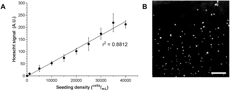
Figure 4.
Hoechst 33342 live cell nuclear staining and fluorescence quantification. C57BL/6J MLFs were serially diluted and plated at various known seeding densities ranging from 1,000 cells/mL to 40,000 cells/mL (n = 8 wells per seeding density) and stained with Hoechst 33342. Fluorescent signal was quantified by microplate reader and shown to be highly correlated to known seeding density (A; linear regression r2 = 0.8812). In B, cells seeded at 40,000 cells/mL and stained with Hoechst 33342 and imaged by fluorescence microscopy. Original magnification 40x; scale bar indicates 100 μm.

