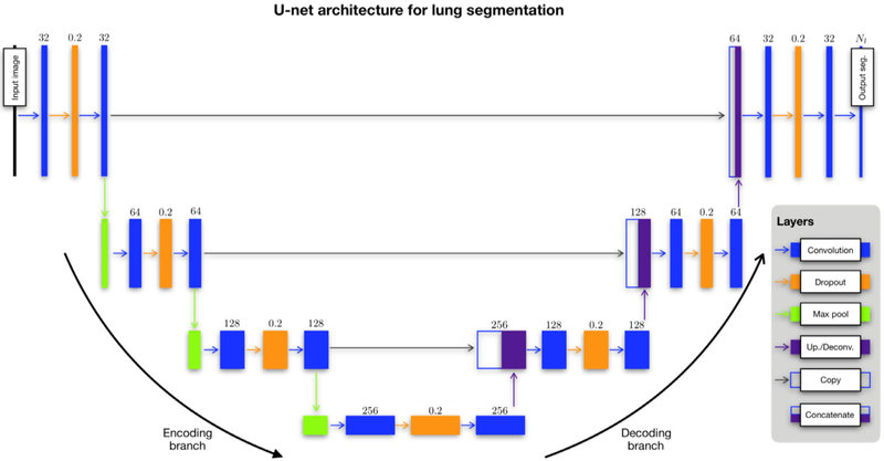
Figure 3.
The modified U-net architecture for both structural and functional lung segmentation (although certain parameters, specifically the number of filters per convolution layer, are specific to the functional case). Network layers are represented as boxes with arrows designating connections between layers. The main parameter value for each layer is provided above the corresponding box. Each layer of the descending (or “encoding”) branch of the network is characterized by two convolutional layers. Modification of the original architecture includes an intermediate dropout layer for regularization (dropout rate = 0.2). A max pooling operation produces the feature map for the next series. The ascending (or “decoding”) branch is similarly characterized. A convolutional transpose operation is used to upsample the feature map following a convolution→ dropout→ convolution layer series until the final convolutional operation which yields the segmentation probability maps.

