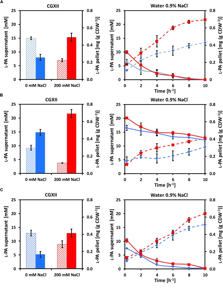
FIGURE 2.
Intracellular (filled columns, straight lines) and extracellular (dashed columns, dashed lines) L-PA concentration profiles of the strains (A) GSL(pVWEx1-lysDH-proC), (B) GSLΔyggB(pVWEx1-lysDH-proC) and (C) GSLΔyggB(pVWEx1-lysDH-proC)(pEKEx3-yggB) after growth in glucose-minimal medium (left panels) without added NaCl (blue) or with 200 mM NaCl (red) and after osmotic downshift to 0.9% NaCl (right panels). Values represent means and standard deviations.

