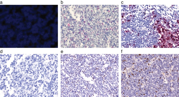
Figure 2.

Detection of Merkel cell polyomavirus (MCPyV) and human polyomavirus 7 (HPyV7) in thymoma ID 1‐8. (a) Magnification (63x) an overlay of nuclei 4,6‐diamidino‐2‐phenylindole dihydrochloride staining and MCPyV fluorescence in situ hybridization of thymoma ID 1‐8 shows that MCPyV DNA is present in thymoma nuclei. Single dots per nuclei were detected. (b) RNAscope RNA in situ hybridization (10x) was performed on the thymoma ID 1‐8, (c) MCPyV‐positive Merkel cell carcinoma (MCC) tissue, and (d) and thymoma ID 1‐32. MCPyV LT RNA are detected in (b) thymoma cells, which is comparable to the dot‐like signals in (c) MCPyV‐positive MCC cells. (d) In thymoma ID‐32, no MCPyV RNA is present. Immunohistochemistry (10x) for (e) MPCPyV with the CM2B4 antibody and (f) HPyV7 with the 2t10 antibody reveal MCPyV and HPyV7 expressing thymoma cells. MCPyV expression is weaker than HPyV7.
