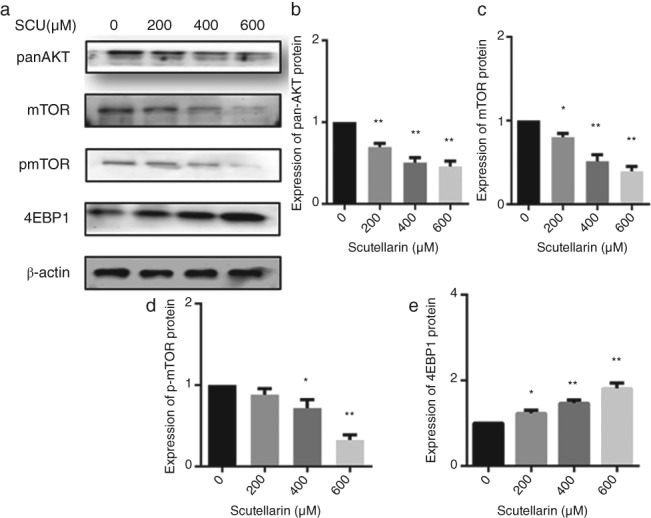
Figure 6.

A549 cells were treated with scutellarin (SCU) (0, 200, 400, 600 μM) for 24 hours. Western blotting analyses were used to detect the expression of proteins in the AKT/mTOR/4EBP1 pathways (a). Data are expressed as means ± standard deviation from three independent experiments. * P < 0.05, ** P < 0.01 vs. the control group (b, c, d, e).
