
Figure 1.
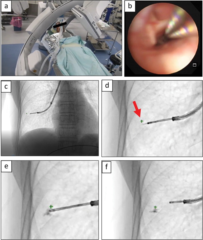
Bronchoscopic video‐assisted thoracoscopic surgery (VATS) marker injection under cone beam computed tomography (CBCT)‐augmented X‐ray fluoroscopy guidance. (a) CBCT is performed to obtain three‐dimensional positions of multiple pulmonary nodules. (b) Bronchoscopic fluorescent marking after introduction of general anesthesia in a hybrid operating room. (c) X‐ray fluoroscopic image with the transbronchial aspiration cytology needle inserted into the peripheral lung. (d) The X‐ray‐invisible pulmonary nodule is indicated as “+” on X‐ray fluoroscopy (red arrow). (e) VATS marker solution is injected near the target. (f) The VATS marker is placed successfully.