Figure 4.
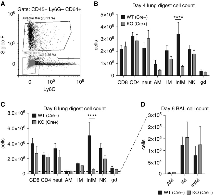
M-TBK1-KO mice have fewer recruited inflammatory lung macrophages during influenza infection. Mice were infected intranasally with 5 × 10⁴ egg-infective dose and lungs were harvested and digested to single-cell suspensions. (A) Representative flow cytometric plot from CD45+/Ly6G−/CD64+ gate showing definition of resident alveolar (AM; Siglec F+/Ly6Clow), resident interstitial (IM; Siglec F−/Ly6C−), and recruited inflammatory (InfM; Siglec F−/Ly6C+) macrophages. Total number of relevant cell populations identified by flow cytometry at (B) Day 4 and (C) Day 6 after infection. (D) Macrophage subpopulations recovered from BAL at Day 6 after infection. n = 6–8 mice per genotype per group and two independent experiments analyzed by Mann-Whitney U test. NK = natural killer cells. ****P < 0.001. gd = γδ.

