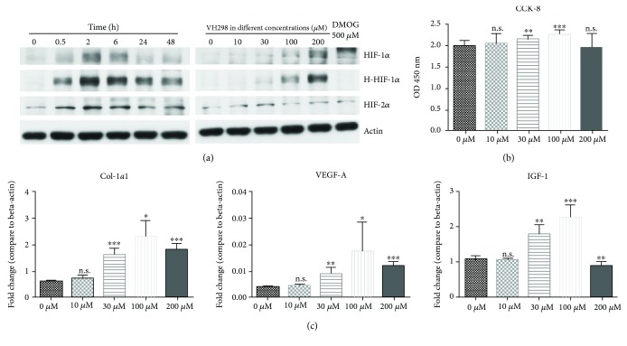
Figure 1.
Protein concentration of HIF-1α, HIF-2α, and hydroxy-HIF-1α, cell proliferation, and multiple gene expressions were related to the VH298 dose. (a) After VH298 or DMOG treatment, protein levels in rFb were detected by immunoblots. Protein concentration of HIF-1α, HIF-2α, and hydroxy-HIF-1α in rFb increased gradually along with VH298 concentration, and DMOG only upregulated protein levels of HIF-1α and HIF-2α, but not hydroxy-HIF-1α, after 6 h treatment. 200 μM VH298 increased the HIF-1α, HIF-2α, and hydroxy-HIF-1α protein levels up to 2 h and was followed by a decrease. (b) Cell viability of rFb was evaluated by the CCK-8 assay. VH298 promoted cell proliferation at doses of 30 μM and 100 μM. Graphs represent mean ± SD (VH298-treated vs. control) (n = 12). (c) Col1-α1, VEGF-A, and IGF-1 gene expressions in rFb were detected by quantitative real-time PCR after treatment with VH298 at different doses, and 30 μM and 100 μM were the most stable doses for upregulation of gene expression. Graphs represent mean ± SD (VH298-treated vs. control) (n = 6). ∗P < 0.05, ∗∗P < 0.01, and ∗∗∗P < 0.001; OD: optical density.

