Figure 3.
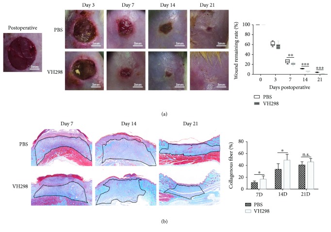
Wound healing rate and collagen accumulation in wound tissue. (a) The remaining wound area was measured after photography. VH298-treated wounds healed much faster compared to PBS-treated wounds. Graphs represent mean ± SD (VH298-treated vs. PBS-treated) (n = 8). (b) Masson trichrome staining showed more collagen deposition (blue) in the granulation tissue (black line-circled area), in which quantitative measurement was applied, and the collagen deposition ratio was significantly increased in the VH298-treated group compared to the PBS-treated group at the early and middle stages (7 days and 14 days). Graphs represent mean ± SD (VH298-treated vs. PBS-treated) (n = 8). ∗P < 0.05, ∗∗P < 0.01, and ∗∗∗P < 0.001. D: day; n.s.: not significant.

