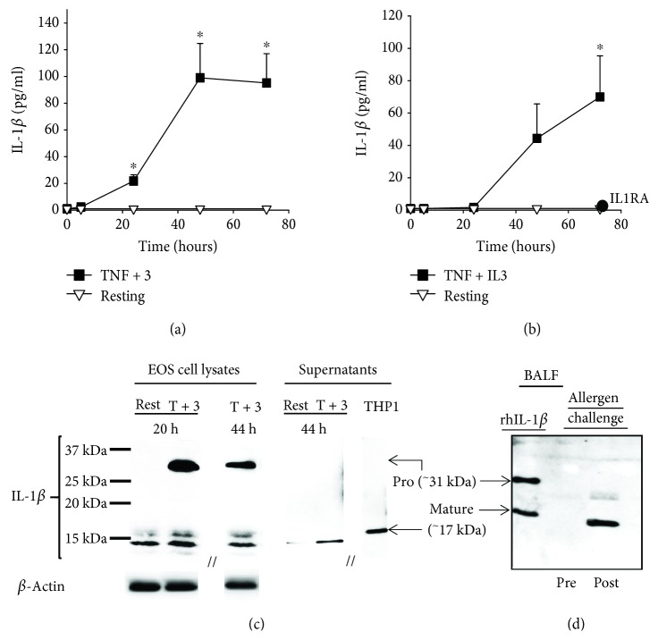
Figure 2.
Activated eosinophils release bioactive IL-1β protein with a MW < 17 kDa. (a and b) Blood eosinophils were cultured with medium alone (resting) or TNF-α plus IL-3 (TNF + 3) for 5, 24, 48, and 72 h. (a) IL-1β present in eosinophil cultures was measured by ELISA. (b) The bioactivity of IL-1β was determined using HEK-Blue™ IL-1β reporter cells and interpolation on a standard curve produced using a recombinant human mature IL-1β. The addition of IL1RA (0.4 μg/ml) to the reporter cells completely inhibited the bioactivity of the conditioned media obtained 72 h after eosinophil stimulation with TNF + IL3. Data are mean ± SEM of experiments on eosinophil preparations from five to six subjects (a) and three subjects (b). ∗ p < 0.05 for TNF-α plus IL-3 versus resting, at the corresponding time point. (c) Blood eosinophils were cultured with medium alone (rest) or TNF-α plus IL-3 (T + 3) for 20 and 44 h. Cell lysates and concentrated conditioned media (supernatants) were prepared. Western blots were performed to visualize IL-1β protein sizes. Supernatant fluid from activated THP-1 were used as a positive control for the 17 kDa mature form of IL-1β. The blot shown is representative of three using three different blood eosinophil donors. (d) BAL fluids (BALF) obtained by bronchoscopy before (Pre) and 48 h after (Post) a segmental allergen challenge were evaluated by western blot for the presence and size of IL-1β. Positive control (rhIL-1β) includes both pro- and mature forms of IL-1β. The blot is representative of two experiments using BAL fluids from two different subjects.

