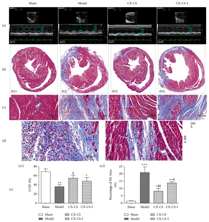
Figure 2.
LVEF, percentage of MI area, and histopathological observations after inhibition of Notch γ secretase. (a) Effect of Notch inhibition on LVEF. (b) Effect of Notch inhibition on percentage of MI area. (c) Histopathological observations after inhibition of Notch. (d) Granulation observation. (e) Statistical analysis of LVEF and percentage of MI area. No. 1 capillaries (arrows point to endothelial cells), No. 2 fibroblast, No. 3 inflammatory cell, No. 4 collagen fibre, No. 5 normal cardiac myocytes, No. 6 hypertrophic cardiomyocyte, and No. 7 dissolution of cardiac myocyte. ∗P<0.05, ∗∗P<0.01, ∗∗∗P<0.001 vs. sham group; $P<0.05, and $$P<0.01 vs. model group. n=6.

