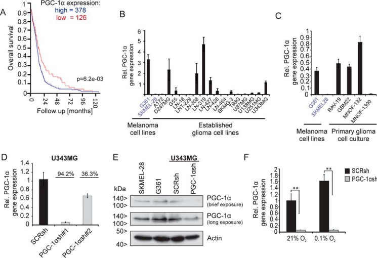
Figure 1.
Knockdown of PGC-1α in the U343MG glioblastoma cell line. A, R2 database analysis shows negative correlation between PGC-1α expression and survival in glioma patients. The investigation was performed using only glioblastoma samples and the dataset “Tumor Glioblastoma–TCGA–540–MAS5.0–u133a” in the R2 database using overall survival data and 1st quartile of gene expression as a cutoff parameter. B and C, established glioma cell lines (B) and primary glioblastoma cell cultures (C) were analyzed for PGC-1α mRNA expression by qPCR. The melanoma cell line G361 served as positive and SKMEL-28 as negative control for PGC-1α expression. D, quantitative qPCR analysis confirms knockdown of PGC-1α in U343MG glioma cell line. E, SKMEL-28, G361, and U343MG SCRsh and PGC-1αsh#1 cells were incubated for 24 h in SFM, and PGC-1α and actin were analyzed by immunoblot. F, PGC-1α expression in SCRsh and PGC-1αsh#1 cells under normoxic conditions (21%) was compared with hypoxic conditions (0.1%), cells were incubated in serum-free medium.

