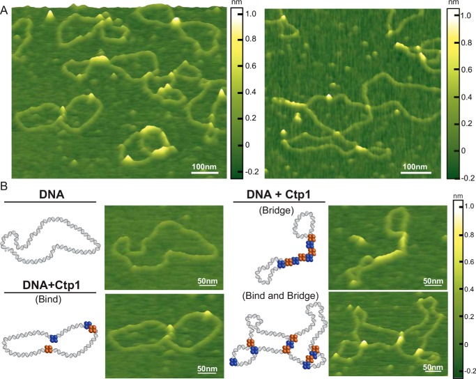
Figure 1.
Ctp1 tetramers form bridging filaments on DNA. A, representative atomic force microscopy images of 800 nm Ctp1 with 1 nm pHOT1 relaxed plasmid DNA. White scale bar, 100 nm. B, selected examples of relaxed plasmid DNA, Ctp1–DNA binding, and Ctp1–DNA bridging complexes by AFM. Both intra- and intermolecular bridging events are shown. Blue and orange spheres, Ctp1 protomers involved in DNA interactions. White scale bar, 50 nm.

