Fig. 8.
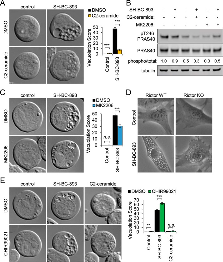
Ceramide inhibits vacuolation by reducing Akt activity. A, FL5.12 cells were treated for 3 h with SH-BC-893 (2.5 μm) and/or C2-ceramide (50 μm) as indicated and imaged by DIC microscopy. B, Western blotting measuring the phosphorylation of the AKT substrate PRAS40 (total and phospho-Thr246) in FL5.12 cells treated for 30 min with SH-BC-893 (5 μm), C2-ceramide (50 μm), and/or MK2206 (1 μm) as indicated. C, FL5.12 cells treated for 3 h with SH-BC-893 (2.5 μm) and/or AKT inhibitor MK2206 (1 μm) as indicated and visualized as in (A). D, Rictor WT and KO murine embryonic fibroblasts (MEFs) treated for 6 h with vehicle or SH-BC-893 (5 μm) then visualized by phase contrast microscopy. E, FL5.12 cells treated for 3 h with SH-BC-893 (2.5 μm) or C2-ceramide (50 μm) and the Gsk3b inhibitor CHIR99021 (10 μm) as indicated; visualized as in (A). In (A, C, E), ≥ 400 cells per condition were analyzed from a total of three independent experiments; using a two-tailed t test to compare either SH-BC-893 or C2-ceramide to the vehicle control (in E) or SH-BC-893 alone to the combination of SH-BC-893 with C2-ceramide (A), MK2206 (C), or CHIR99021 (E), n.s. = not significant, ** = p ≤ 0.01, or *** = p ≤ 0.001.

