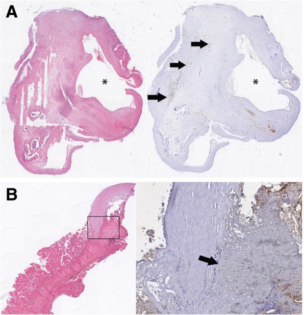
Fig. 2.

a Hematoxylin and eosin (HE) staining (demonstrating the absence of IPH) and the corresponding CD 31 staining (black arrows show presence of microvessels) from a histological specimen obtained during carotid endarterectomy. b Hematoxylin and eosin (HE) staining (demonstrating IPH) and the corresponding CD 31 staining (no presence of microvessels) from a histological specimen obtained during carotid endarterectomy, the black arrow points towards the area were intraplaque hemorrhage is present
