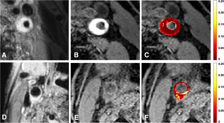
Fig. 3.
Pre-contrast T1 weighted (T1w) quadruple inversion recovery (QIR) turbo spin echo (TSE) image (a) from a patient with intraplaque hemorrhage (IPH). Note that a Regional Saturation Technique (REST) slab is visible on the right side, which was placed over the subcutaneous fat tissue to prevent ghosting artefacts. Three-dimensional T1w inversion recovery turbo field echo (IR TFE) image (b) from the same patient with IPH. A hyperintense signal is visible within the bulk the plaque compared with the adjacent sternocleidomastoid muscle (*), indicating the presence of IPH. Parametric Ktrans map of the plaque is overlaid on IR TFE image shown in B (c). In this parametric map voxelwise determined Ktrans values are colour encoded from 0 to 0.25 min− 1. Within this plaque, the IPH exhibits low Ktrans values, shown in dark red, while higher Ktrans values (brighter red) are observed in the outer vessel wall (adventitial layer). Pre-contrast T1w QIR TSE image from a patient without IPH (d). 3D T1w IR TFE image (e) from the same patient without IPH. Parametric Ktrans map is overlaid on IR TFE image shown in B (f). In this parametric map voxelwise determined Ktrans values are colour encoded from 0 to 0.25 min− 1. Within this plaque, higher Ktrans values are observed, shown in bright red/yellow/white. Written informed consent for publication of their clinical details and/or clinical images was obtained from the patients. A copy of the consent forms is available for review by the Editor of this journal

