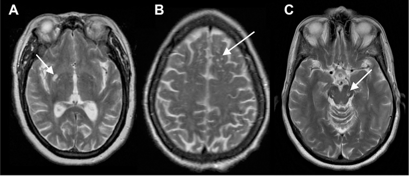
Fig 1.

Characteristic locations of PVSs seen on T2 MR images. Type I: basal ganglia along the course of lenticulostriate arteries (A). Type II: subcortical white matter in the high convexities (B). Type III: midbrain at the pontomesencephalic junction (C).
