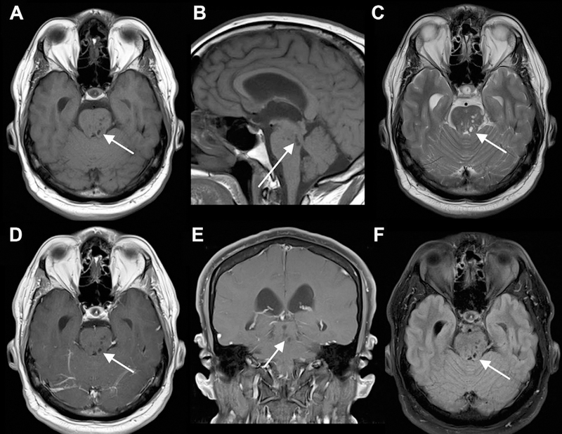
Fig 10.

Minimally dilated PVS causing hydrocephalus. Small dilated PVSs in the midbrain (left more than right) and pons seen on axial and sagittal T1 (A, B), axial T2 (C), axial and coronal T1 postcontrast (D, E), and axial FLAIR (F) MR images. The PVSs are isointense to CSF on all sequences. The lateral and third ventricles are dilated, consistent with compensated, obstructive hydrocephalus.
