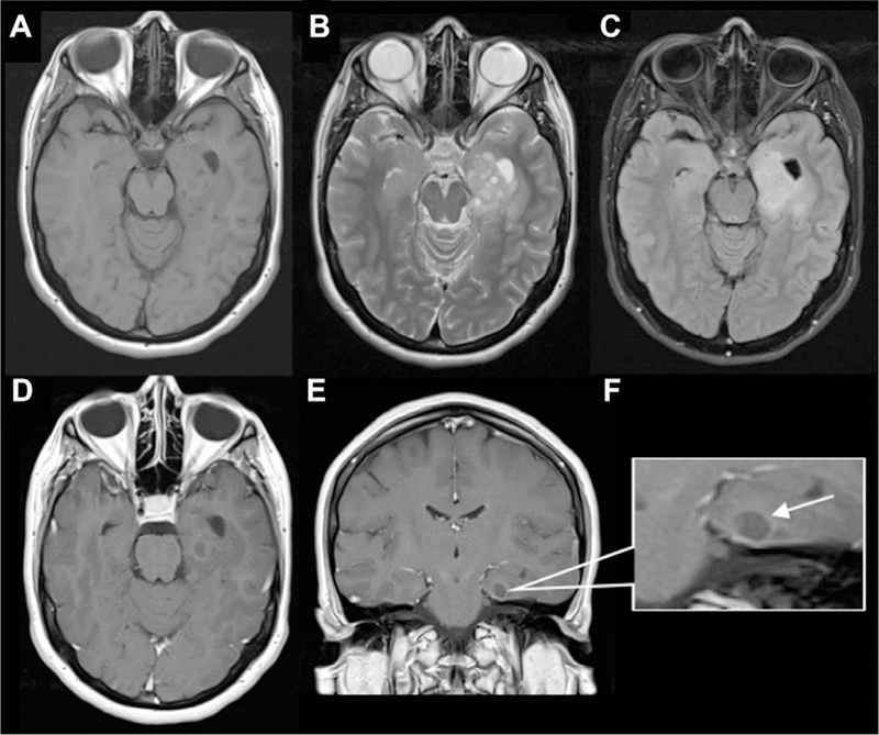
Fig 12.

Dysembryoplastic neuroepithelial tumor (DNET) mimicking dilated PVS. There are numerous small- to medium-sized cystic lesions with “bubbly appearance” involving the amygdala, anterior hippocampus, and anterior parahippocampal gyrus are seen on (A) axial T1 (B) and axial T2 images. There is a larger area of surrounding expansile FLAIR signal abnormality (C) extending through the left posterior hippocampus, mid-fusiform gyrus, and temporal periventricular white matter along the inferolateral temporal horn. Faint peripheral enhancement around one of cystic components in the left parahippocampal gyrus on axial and coronal T1 postcontrast images (D, E).
