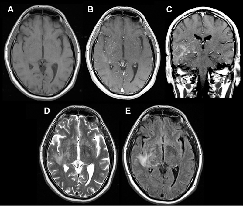
Fig 18.

Sample case of intravascular CNS lymphoma involving PVSs, seen on axial T1 (A), axial and coronal T1 postcontrast (B, C), and axial T2/FLAIR images (D, E). There is linear, predominantly perivascular enhancement extending from the right thalamus and basal ganglia into the corona radiata, frontal, parietal, and temporal lobes with associated T2/FLAIR signal abnormality.
