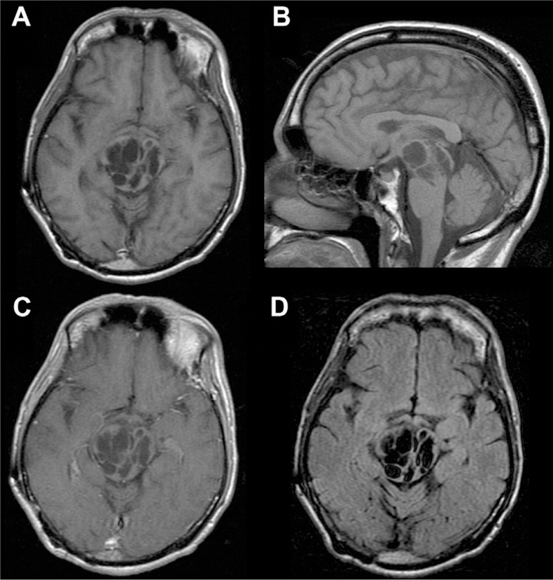
Fig 8.

Giant/tumefactive PVSs. Multiple dilated cystic foci seen in the pons and midbrain extending into the right thalamus with CSF signal intensity consistent with a giant/tumefactive PVS as seen on axial and sagittal T1 (A, B), axial T1 postcontrast (C), and axial FLAIR (D) MR images.
