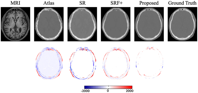
Fig. 11.

First row shows visual comparison of the MR image, the four estimated CT images by other three competing methods and our proposed method, and the ground-truth CT for a typical brain case. The second row shows difference maps between each estimated target CT and the ground-truth CT.
