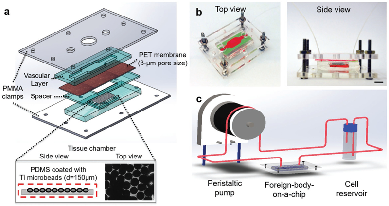
Figure 1.
Design of the FBROC device. a) Exploded schematic diagram showing the multilayer structure of the bioreactor, where an endothelialized porous membrane is sandwiched in between a vascular channel on top and a tissue chamber at the bottom, the latter of which implant of Ti microbeads was placed. b) Perspective- and side-view photographs showing the bioreactor in the multilayer configuration. c) Schematic diagram showing the operation of the FBROC device, where immune cells are circulated from the top vascular channel of the bioreactor to probe their interactions with the Ti microbeads in the bottom tissue chamber through the endothalial barrier.

