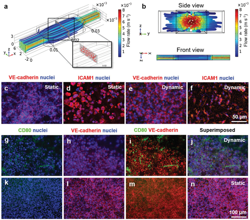
Figure 2.
Characterization of monocyte distribution, vascular barrier, and monocyte-endothelium interactions. a,b) Simulated distributions of the circulating immune cells in the top vascular channel of the bioreactor. c–f) Immunostaining of VE-cadherin and ICAM for confluent HUVECs cultured under (c,d) static and (e,f) dynamic conditions on the porous PET membrane. g–n) THP-1 monocyte interactions with the confluent endothelium under static and dynamic conditions.

