Figure 3.
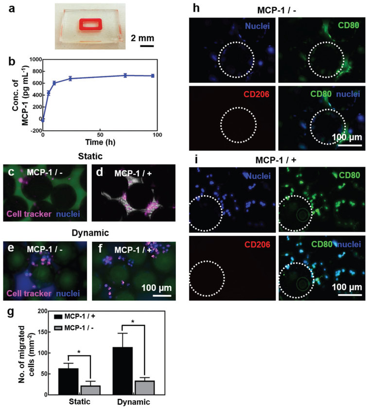
FBR of THP-1 monocytes to the Ti microbeads. a) Photograph showing the GelMA hydrogel ring in the bottom tissue chamber for MCP-1 release. b) MCP-1 release over a 96-h period. c–f) THP-1 monocyte trans-endothelial migration towards the bottom Ti microbeads under (c,d) static and (e,f) dynamic conditions, in the (c,e) absence and (d,f) presence of MCP-1. The cells were pre-labeled with cell tracker (pink) and post-labeled for nuclei (blue). g) Quantifications of the number of THP-1 monocyte migration. h,i) CD80 (green)/CD206 (red) expressions of activated THP-1 monocytes on the Ti microbeads, in the h) absence and i) presence of MCP-1, under dynamic conditions. The nuclei were counterstained in blue. The white dotted circles indicate the Ti microbeads. *p< 0.05.

