Fig. 1.

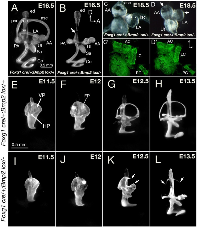
Phenotypes of Foxg1cre/+; Bmp2lox/− inner ears. (A,B) Paint-filled Foxg1cre/+; Bmp2lox/+ (A) and Foxg1cre/+; Bmp2lox/− (B) inner ears at E16.5. (B) The conditional mutants are missing all three semicircular canals, and the common crus is thinner (arrow); however, the endolymphatic duct, utricle, saccule and cochlear duct appear normal. (C,D) Dissected membranous labyrinths of the utricle and the anterior and lateral ampullae in heterozygous controls (C) and in conditional mutants (D) at E18.5. Mutant ampullae have no canal opening (D, arrows) but the cristae within appear intact based on phalloidin staining of sensory hair cells (C′,D′). (E-L) Semicircular canal development in Foxg1cre/+; Bmp2lox/+ (E-H) and Foxg1cre/+; Bmp2lox/− (I-L) ears between E11.5 and E13.5. (E-H) The vertical and lateral canal pouches in heterozygous controls are apparent at E11.5, with fusion plates emerging by E12 and followed by resorption. Canals reach their adult pattern by E13.5, but the size of the canals continues to increase after this age. (I-L) The Foxg1cre/+; Bmp2lox/− canal pouch (I) is slightly smaller than controls (E) at E11.5, but reduction in size is clear by E12 (J). (K) At E12.5, an opening is observed in the anterior region of the vertical canal pouch (arrows). (L) By E13.5, only remnants of the three canals are evident (arrows). AA, anterior ampulla; AC, anterior crista; asc, anterior semicircular canal; CC, common crus; Co, cochlea; ed, endolymphatic duct; FP, fusion plate; HP, horizontal canal pouch; LA, lateral ampulla; LC, lateral crista; lsc, lateral semicircular canal; PA, posterior ampulla; PC, posterior crista; psc, posterior semicircular canal; VP, vertical canal pouch; Sa, saccule; Ut, utricle. Orientations: A, anterior; D, dorsal. Orientation in B also applies to A,E-L. Orientation in D′ applies to C-D. Scale bars: 0.5 mm in A for A,B; 0.5 mm in E, for E-L.
