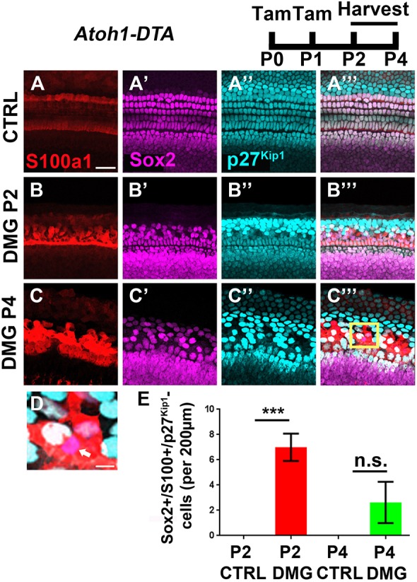
Fig. 6.

p27Kip1 is specifically downregulated among PCs/DCs during spontaneous HC regeneration in the neonatal mouse cochlea. (A-C‴) Representative confocal slice images with anti-S100a1 (red), anti-Sox2 (magenta), and anti-p27Kip1 (cyan) antibodies used to investigate changes in the expression of the cell cycle inhibitor p27Kip1 in SCs after HC damage (DMG). Controls (CTRL) for each time point were littermates that lacked either the Atoh1-CreERTM or Rosa26DTA allele (A-A‴). Atoh1-DTA mice were injected with tamoxifen (Tam) at P0/P1 to induce HC death and cochleae were collected at P2 or P4 (B-C‴). All Sox2-positive/p27Kip1-negative cells observed were also S100a1-positive, which suggests that they were PCs and DCs. Scale bar: 25 µm. (D) Higher magnification image of the boxed region in C‴ showing a Sox2-positive/p27Kip1-negative cell (white arrow). Scale bar: 5 µm. (E) A small number of Sox2-positive/S100a1-positive/p27Kip1-negative PCs/DCs were detected at P2 and P4 after HC damage, whereas no p27Kip1-negative cells were detected in controls at any age or outside the S100a1 region. ***P<0.001, determined using a one-way ANOVA with a Sidak's post-hoc test. Data are mean±s.e.m.; n=3-5. n.s., not significant.
