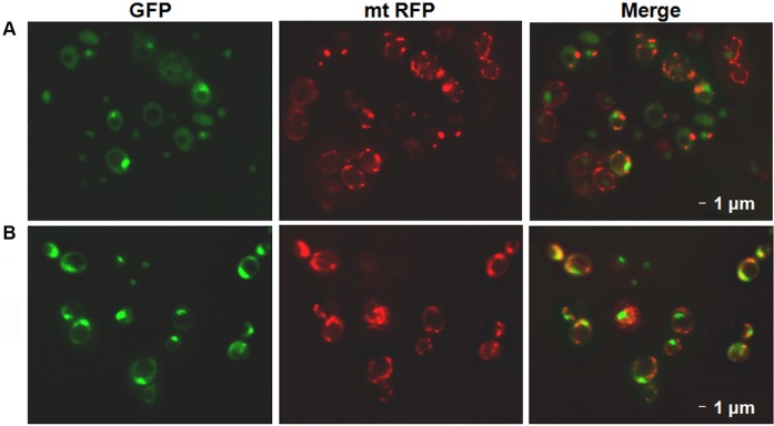
Fig. 5.
GFP fluorescence colocalization of Gcn5. (A,B) Fluorescence microscopy of the GCN5-GFP with the endogenous GCN5 fused with the GFP gene grown in YP 2% glucose (A) or 3% glycerol (B) containing media to stationary phase. The fluorescent signals of the GCN5-GFP clone transformed with pmtRFP plasmid overexpressing the mitochondrial Red Fluorescent Protein show colocalization between Gcn5 and mitochondria.

