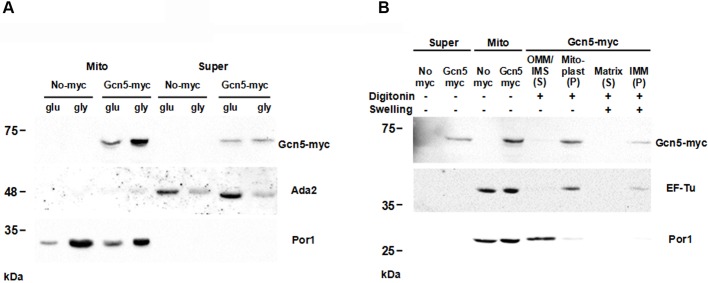
Fig. 6.
Subcellular and submitochondrial fractions indicate that Gcn5 protein is localized in the mitoplasts. (A) Whole cell lysate (Super) and mitochondrial (Mito) fractions of W303-1A GCN5-9Myc or untagged control (No-myc) strains grown in YP 2% glucose or 3% glycerol containing media were analysed by 10% SDS PAGE. Gcn5-myc, Ada2 and Por1 antibodies were used as nuclear and mitochondrial marks, respectively. (B) Purified mitochondria (Mito) and supernatant (Super) from rho+ cells were treated (+) or not (−) with Digitonin or swelling buffer and sonication (Swelling) to obtain mitochondrial fractions. After isolation of outer membrane and inner membrane space (OMM/IMS), mitoplasts were fractionated in inner membrane (IMM) and matrix. Pellet (P) and soluble (S) fractions were isolated by ultra centrifugation and immunostained with antibodies against the Gcn5-myc, EF-Tu and Por1 for marks of different mitochondrial compartments (see Materials and Methods for details).

