Fig. 2.
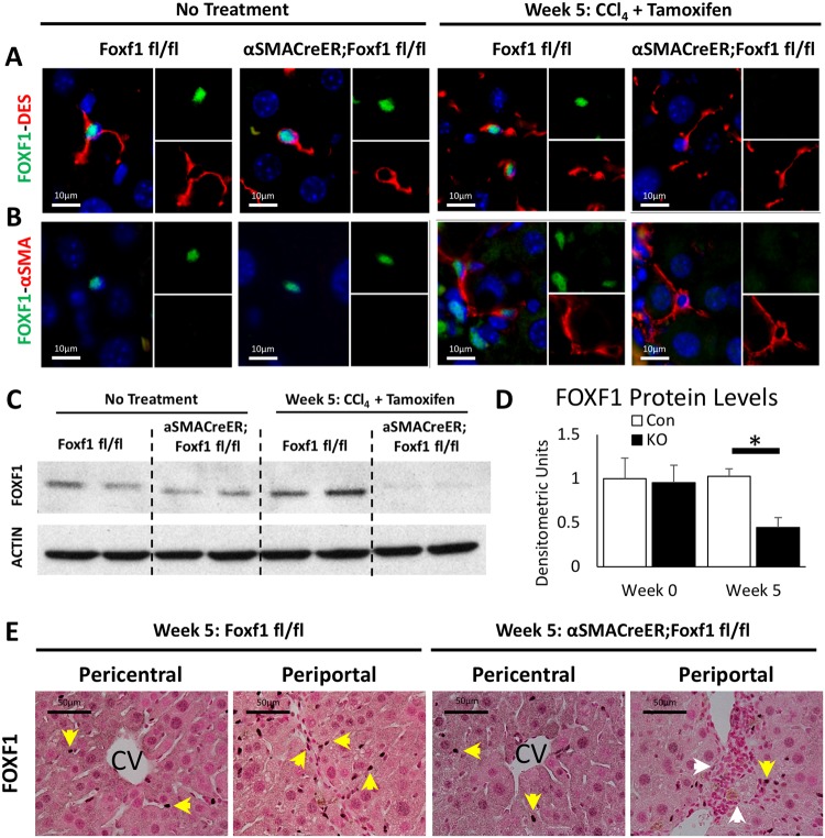
αSMA-CreER effectively deletes Foxf1 from hepatic myofibroblasts. (A,B) FOXF1 co-localizes with DES in HSCs before and after CCl4-induced injury. FOXF1 co-localizes with DES and αSMA in MFs after chronic liver injury. αSMA-CreER effectively deletes Foxf1 from MFs after Tam treatment. (C) Western blot shows total liver protein levels of FOXF1 are decreased in αSMACreER;Foxf1−/− livers after CCl4 injury. Cropped blots are presented here with full length blots presented in Fig. S12. (D) Quantification of western blot revealed a significant loss of FOXF1 in αSMACreER;Foxf1−/− livers. Quantification was averaged across four blots. FOXF1 levels were internally normalized to ACTIN for each sample. *P<0.05. (E) FOXF1 staining is detected in liver parenchyma and fibrotic regions (yellow arrows). FOXF1 staining is decreased in fibrotic regions of αSMACreER;Foxf1−/− livers (white arrows).

