Fig. 1.
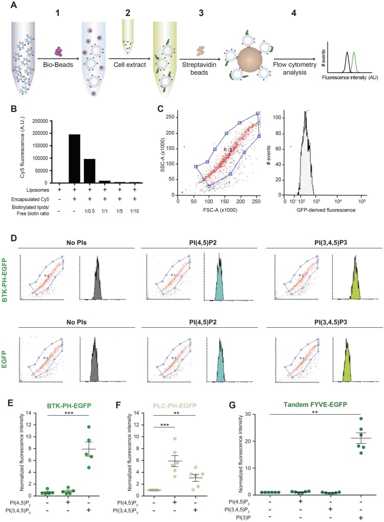
ProLIF is a flow cytometry-based assay for detection of specific protein-lipid interactions. (A) Outline of ProLIF workflow. Step 1: Bio-Beads™ are added to lipids solubilised in Triton X-100 to remove the detergent and obtain liposomes. Step 2: liposomes are incubated with membrane-free cell extract containing the EGFP-tagged protein of interest. Step 3: Streptavidin–Sepharose (SA) beads are added in order to capture the liposomes via interaction with biotinylated lipids present in the liposome membrane. Step 4: SA beads are analysed by flow cytometry (FACS). Red dots and blue dots represent biotinylated lipids and PIs, respectively. Green fragments represent EGFP-tagged proteins from the cell lysate. (B) Biotinylated-lipid-containing liposomes were generated with and without encapsulated Cy5 dye, captured on SA beads in the presence or absence of increasing amounts of free biotin and analysed via FACS. The molar ratio between biotinylated lipids and soluble biotin added in each sample is indicated (n=1). (C) Scatter plot and fluorescence histogram from SA beads alone incubated with cell lysate from EGFP-transfected cells and analysed by FACS. (D) Biotinylated-lipid-containing liposomes, with the indicated PI content, were incubated with cell lysates from EGFP alone- or BTK-PH–EGFP-transfected cells (equal EGFP concentrations) and then captured by SA beads and analysed by FACS. Shown are representative dot blots, and size gating in FACS, and histograms depicting EGFP fluorescence intensity (FL1) profiles (note that the axis labels are as in C). The red population in the scatter plot was gated for quantification. Data shown represent three individual experiments. (E) Binding of the BTK-PH–EGFP domain (from cell lysate as in D) to biotinylated-lipid-containing liposomes, with the indicated PI content, relative to control PI-free liposomes (data are normalised median fluorescence intensities shown as the mean±s.e.m.; n=5 independent experiments). (F) Binding of EGFP-tagged PLC-PH domain (from cell lysate) to biotinylated-lipid-containing liposomes, with the indicated PI content, relative to control PI-free liposomes (data are normalised median fluorescence intensities shown as the mean±s.e.m.; n=5 independent experiments). (G) Binding of tandem FYVE-EGFP domains (from cell lysate) to biotinylated-lipid-containing liposomes, with the indicated PI content, relative to PI-free liposomes (data are normalised median fluorescence intensities shown as the mean ±s.e.m.; n=6 independent experiments). **P<0.01, ***P<0.001

