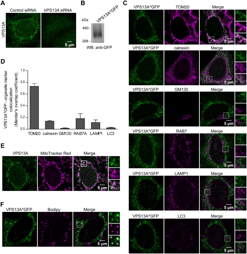
Fig. 5.
VPS13A preferentially colocalizes with mitochondria. (A) Endogenous VPS13A in HeLa cells was labeled by immunofluorescence and visualized by confocal microscopy. VPS13A presents a distinct subcellular network-like pattern that disappears upon treatment with VPS13A siRNAs. Images are representative of three independent experiments. (B) VPS13A was tagged with GFP at an internal position and lysates of HeLa cells transiently expressing the resulting VPS13A^GFP protein were analyzed by western blotting using an anti-GFP antibody to verify that the tagged protein had the expected molecular size. (C) Transiently transfected VPS13A^GFP HeLa cells were fixed and labeled with antibodies against TOM20 (also known as TOMM20), calnexin, GM130 (also known as GOLGA2), RAB7A, LAMP1 or LC3B (markers of mitochondria, the ER, cis-Golgi, endosomes, lysosomes or autophagosomes, respectively). Examples of VPS13A^GFP puncta colocalizing with those markers are enlarged in the selected areas. Representative images of two independent experiments are shown. (D) Quantification of the colocalization of VPS13A with the above-mentioned organelle markers from an average of 13 cells per marker. The means±s.d. of Mander's overlap coefficient for VPS13A after thresholding are plotted. (E) Endogenous VPS13A was labeled by immunofluorescence and visualized in HeLa cells previously treated with Mitotracker Red CMX ROS to stain mitochondria. (F) Transiently transfected VPS13A^GFP HeLa cells were treated with BODIPY to stain lipid droplets. Enlargements of selected areas are shown to better visualize the colocalization between VPS13A and the organelle markers.

