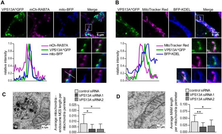
Fig. 6.
VPS13A localizes in regions in which mitochondria overlap with other organelles and can influence the length of mitochondria in contact with other organelles. (A,B) VPS13A^GFP was visualized in cells transiently transfected with mCherry-RAB7A and mito-BFP to label endosomes and mitochondria (A) or cells transiently transfected with BFP-KDEL and stained with Mitotracker Red CMX ROS to label the ER and mitochondria (B). Images are representative of 10 cells. Enlargements of regions in which mitochondria overlap with endosomes (A) or the ER (B) are also shown. For a better visualization of VPS13A localization at the interface between organelles, the plots represent the relative intensity of the fluorescent signal for each marker along the depicted lines in the enlarged regions. (C,D) HeLa cells treated with control or VPS13A siRNAs were observed by TEM. MCS between mitochondria and endosomes (C) and between mitochondria and the ER (D) were identified (an example of each type of MCS is shown) and their average length per mitochondrial perimeter was measured in 17 random cell sections per sample. MAM, mitochondria-associated ER membrane. Means±s.d. are plotted (*P<0.05, **P<0.01).

