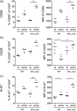
Figure 2.

Activation status of CD4+ T cells in mouse cytomegalovirus (MCMV)‐infected wild‐type (WT) and interferon (IFN)‐γ knock‐out (KO) mice. Percentage of CD69+, CD25+ or Ki‐67+CD4+ T cells (a–c, left panel), gated as % CD69+, CD25+ or Ki‐67+ of live singlet CD4+ lymph node cells and their median fluorescence intensity (MFI) (a–c, right panel). Data obtained on day 5 post‐infection in the inguinal lymph nodes. Dots represent individual animals. Horizontal bars refer to median values. NI = not infected. *P < 0·05; **P < 0·01; ***P < 0·001; Kruskal–Wallis with Dunn's post‐test. Depicted data are from one experiment and representative of four to 10 independent experiments with at least five mice per group.
