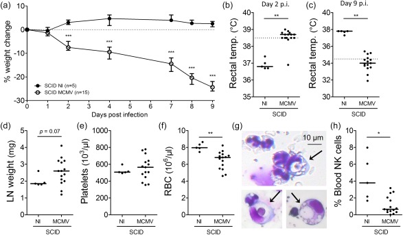
Figure 4.

Severe combined immunodeficient (SCID) mice, lacking functional T and B cells, develop a haemophagocytic lymphohistiocytosis (HLH)‐like syndrome post‐infection with 5 × 10³ plaque‐forming units (pfu) of mouse cytomegalovirus (MCMV). (a) Percentage change in body weight relative to initial weight at day 0 post‐infection (p.i.). Median with interquartile range of five to 15 mice per group. (b,c) Rectal body temperature (temp.) (°C) at days 2 and 9 p.i. Dotted line = 38·5°C (fever) or 34·5°C (end‐point as indication of mortality). (d) Weight of both inguinal lymph nodes (LN) (mg). (e,f) Absolute platelet and red blood cell count in whole blood. (g) Haemophagocytes detected in haematoxylin and eosin (H&E)‐stained cytospins from blood leucocytes of MCMV‐infected SCID mice. Arrows indicate engulfed cells. (h) Natural killer (NK) cell percentage in blood, gated as CD122+CD49b+ cells of CD3– propidium iodide (PI)– leucocytes. (b–f,h) Dots represent individual animals. Horizontal bars refer to median values. (c–h) Data were obtained on day 9 p.i. NI = not infected; *P < 0·05; **P < 0·01; ***P < 0·001; Mann–Whitney U‐test. Depicted data are from one experiment and representative of two experiments with at least five mice per group. [Colour figure can be viewed at wileyonlinelibrary.com]
