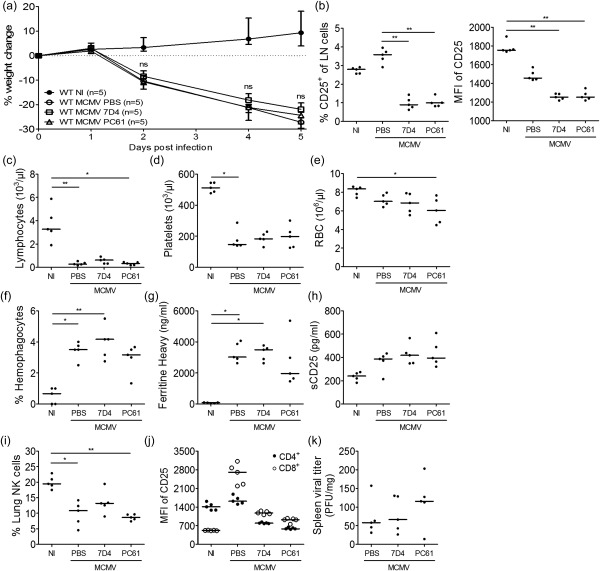
Figure 6.

CD25‐targeting therapy does not ameliorate the haemophagocytic lymphohistiocytosis (HLH)‐like syndrome in mouse cytomegalovirus (MCMV)‐infected wild‐type (WT) BALB/c mice. Anti‐CD25‐antibodies (blocking clone 7D4 or depleting clone PC61) or phosphate‐buffered saline (PBS) were injected intraperitoneally on day 2 post‐infection (p.i.). (a) Percentage change in body weight relative to initial weight at day 0 p.i. Median with interquartile range of five mice per group. (b) Percentage CD25‐expressing cells of live singlet inguinal lymph node (LN) cells (left panel) and their median fluorescence intensity (MFI) of CD25. Clone 3C7 was used to detect remaining CD25 expression. (c–e) Absolute lymphocyte, platelet and red blood cell (RBC) count in whole blood. (f) Percentage haemophagocytes detected in cytospins of leucocytes derived from lung. Dots represent the average of triplicate counts of 100 cells from one mouse. (g) Plasma concentration of the ferritin heavy chain (ng/ml). Dots represent the average of two dilutions for one mouse. (h) Plasma concentration of sCD25 (pg/ml). (i) Natural killer (NK) cell percentage in lung, gated as CD122+CD49b+ cells of CD3– ZombieAqua– singlet cells. (j) MFI of CD25 on CD4+ and CD8+ T cells in the inguinal lymph nodes. Clone 3C7 was used to detect remaining CD25 expression. (k) Titre of infectious virus in spleen [plaque‐forming units (pfu) per mg tissue]. (b–k) Data obtained on day 5 p.i. (b–e, h–k). Dots represent individual animals. Horizontal bars refer to median values. NI = not infected; ns = not significant (P ≥ 0·05); *P < 0·05; **P < 0·01; ***P < 0·001; Kruskal–Wallis with Dunn's post‐test. Depicted data are from one experiment.
