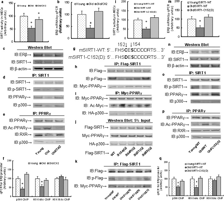
Figure 3.

Reduced ERβ expression in the endothelium from aging mice is due to compromised phosphorylation of amino acid S154 in SIRT1, and the mutant SIRT1‐C152(D) restores this effect. (a–f) The MECs from either Young or Old mice were infected by either control or CK2 lentivirus, and the cells were harvested for further analysis. (a) SIRT1 activity. (b) ERβ mRNA. (c) Protein analysis by Western blotting. (d) IP/WB analysis for SIRT1. (e) IP/WB analysis for PPARγ. (f) ChIP analysis on ERβ promoter. n = 4. *, P < 0.05, vs. Young group. (g) Schematic model for mouse single‐mutant mSIRT1‐C152(D). (h–j) The conditionally immortalized Young MECs were transfected by either Flag‐SIRT1 WT (wild‐type) or single mutants for further analysis. (h) IP/WB analysis of Flag‐SIRT1. (i) IP/WB analysis of Myc‐PPARγ. (j) Western blots analysis of 5% input. (k) The conditionally immortalized MECs from Young or Old mice were transfected by either Flag‐SIRT1 WT (wild‐type) or single mutants for IP/WB analysis. (l–q) The MECs from either Young or Old mice were infected by either SIRT1‐WT or single‐mutant C152(D) lentivirus, and the cells were harvested for further analysis. (l) SIRT1 activity. (m) ERβ mRNA. (n) Protein analysis by Western blotting. (o) IP/WB analysis for SIRT1. (p) IP/WB analysis for PPARγ. (q) ChIP analysis on ERβ promoter. n = 4. *, P < 0.05, vs. Young group. Results are expressed as mean ± SEM.
